The Collateral Ligaments: Your Knee's Side-to-Side Guardians
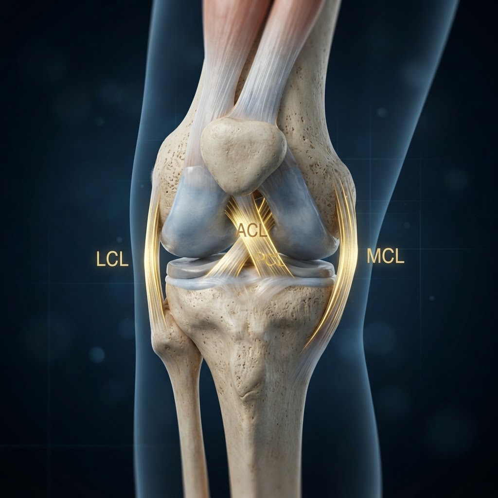
While the ACL gets most of the attention, the medial collateral ligament (MCL) and lateral collateral ligament (LCL) are equally critical for athletic performance. These paired structures control valgus (inward) and varus (outward) forces at the knee — the exact forces generated during cutting, turning, and side-stepping.
Anatomy of the MCL
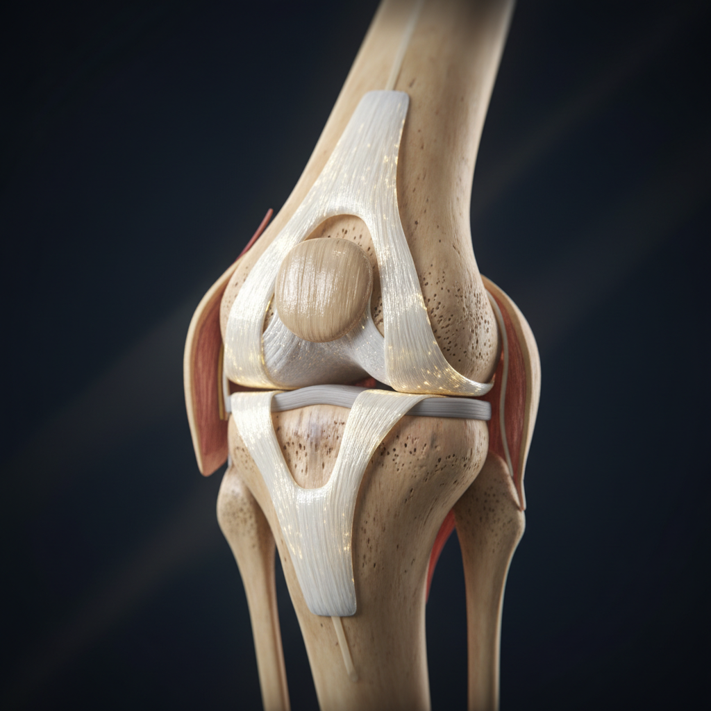
The MCL runs along the inner side of the knee from the femur to the tibia. It has two layers:
- Superficial MCL — the primary restraint against valgus stress
- Deep MCL — merges with the knee capsule, provides secondary support
The MCL also resists external rotation of the tibia, making it essential for the rotational stability needed in sports like football, GAA, and hurling.
Anatomy of the LCL
The LCL runs along the outer side of the knee from the femur to the fibula. Unlike the MCL, it is a discrete cord-like structure separate from the joint capsule. It resists varus stress and works with the popliteus tendon and posterolateral corner to control external rotation.
Why Collateral Ligaments Matter for Cutting and Turning
Every time an athlete changes direction, the knee experiences a combination of:
- Valgus or varus load — the knee is pushed inward or outward
- Rotational force — the body turns while the foot is planted
- Ground reaction force — body weight plus momentum
The collateral ligaments must absorb and manage these forces. If they are weak or poorly conditioned, the knee collapses inward (valgus collapse) — a primary mechanism for both ACL and MCL injuries.
Common Injury Mechanisms
MCL injuries typically occur from:
- A direct blow to the outside of the knee (common in rugby and football)
- Valgus collapse during cutting or landing
- Combined rotational forces
LCL injuries are less common but occur from:
- Varus force to the knee (blow to the inside)
- Hyperextension with rotation
- Often part of a complex posterolateral corner injury
Enhancing Collateral Ligament Performance
Unlike the ACL, the MCL has excellent healing capacity due to its rich blood supply. Both collateral ligaments respond well to progressive loading:
- Isometric loading builds baseline tensile strength
- Controlled lateral movement trains the ligaments to manage side-to-side forces
- Proprioceptive work improves the knee's ability to detect and correct dangerous positions
- Integrated chain training ensures the hip and ankle support the knee's collateral system
The I Am 900 Approach
Our sensor system records the loading response of the collateral system during lateral movement. By mapping valgus and varus patterns during cutting and turning, we identify where the knee's lateral response can be examined — and the effect of any change is compared in a retest.
The collateral ligaments are the knee's foundation for every sidestep, pivot, and turn. Understanding and conditioning them is essential for any athlete whose sport demands lateral movement.
Sport Application — Lateral Movement / Change of Direction
This is a measurement example, not a case study.
For a lateral movement task, the relevant measurement questions are:
- How is pressure distributed?
- What changes during lateral loading?
- Does timing change?
- Does movement sequence change?
- Is the response repeatable?
- Does the athlete produce the same response after calibration?
The sensor system records the loading response during these tasks. It does not directly measure ligament force.
Each question is answered by comparison — baseline, change the input, retest the same defined task, compare what changed.
What Should You Measure in Sport? sets out the measurement vocabulary in full.
Keep Exploring
For the measurement layer of this topic — what can be recorded during a lateral movement task — see MCL and LCL in Lateral Movement: Measuring the Athlete's Response.